
|
November 14, 2006 |
Speak, Memory: Research Challenges Theory of Memory Storage
During sleep, freshly minted memories move from the hippocampus, part of the “old” brain, to the neocortex, or “new” brain, for long-term storage. This has been the reigning theory for decades. Brown University research provides the strongest proof yet of this interaction between the old and new brains – and offers surprising evidence that challenges critical details of this theory of learning and memory. Results appear in Nature Neuroscience. | |||
|
Brown University Home |
PROVIDENCE, R.I. — Daily events are minted into memories in the hippocampus, one of the oldest parts of the brain. For long-term storage, scientists believe that memories move to the neocortex, or “new bark,” the gray matter covering the hippocampus. This transfer process occurs during sleep, especially during deep, dreamless sleep. Many neuroscientists have embraced and built upon this theory of memory storage, or consolidation, for a generation. But the theory is difficult to test. New research led by Brown University neuroscientist Mayank Mehta, conducted with Nobel Prize-winning physiologist Bert Sakmann, shows the best evidence yet of the sleep dialogue between the old brain and the new. 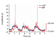 While you were out ... Their work, published in Nature Neuroscience, also shows that this interaction occurs in a startling way. Instead of the hippocampus uploading information to the neocortex in a burst of brain cell communication, Mehta found the opposite: the neocortex seems to drive the dialogue with the hippocampus. The findings may give scientists a new understanding of how the brain manages memories in health and during dementia, offering up a fresh look at the causes of diseases such as Alzheimer’s, as well as potential treatments. “Long-term memory making may be a very different process than we previously thought,” said Mehta, an assistant professor in the Department of Neuroscience at Brown. “Either this reversed dialogue is, somehow, a part of memory storage or this transfer of information from the old to the new brain may not occur during sleep. Either way, the results call into question commonly held theories about the role of cortico-hippocampal dialogue in sleep.” Edvard Moser, a professor at the Norwegian University of Science and Technology and director of the Centre for the Biology of Memory, is a leading expert on memory processes in the hippocampus. Moser said of the new research, “This technically sophisticated study may significantly influence our view of hippocampal-neocortical interactions during sleep-related memory consolidation processes.” To study this dialogue, Mehta recorded electrical activity in rat brains. To mimic the deepest sleep states, the rats were anesthetized then fitted with two electrodes. One electrode measured the electrical activity of thousands of cells in the neocortex. These cells were excitatory, meaning that they spark communication between nerve cells. The other electrode recorded the activity of a single inhibitory cell in the hippocampus. These inhibitory cells shut down dialogue between nerve cells. Using this groundbreaking single-cell recording technique, honed in Sakmann’s laboratory at the Max Planck Institute for Medical Research, researchers made an important finding: During deepest sleep, in both the hippocampus and neocortex, the patterns of neural activity are both regular and highly related. The cells in the old and new brains fired nearly in synch, evidenced by similar peaks and troughs shown on electroencephalographs. This is surprising; Previous studies showed that during deep sleep, when the excitatory cells in the neocortex showed rhythmic activity, excitatory neurons in the hippocampus showed erratic activity. This stumped Mehta and his colleagues: If these two parts of the brain talk during deep sleep, why didn’t they appear to be speaking the same language? They are, Mehta and his team discovered, if you listen to inhibitory, not excitatory, cells in the hippocampus. Mehta and his team also showed that the activity of the cells is related. The timing of activity, or talk, was the same in both brain regions, with a small delay in the hippocampus – as if those cells were echoing the speech in the neocortex. This discovery of synchronized communication between the old and the new brain – a phenomenon known as “phase-locking” – has two key implications. It suggests that the neocortex, not the hippocampus, drives the discussion between these brain systems during deep sleep. It also suggests that the inhibitory neurons control the conversation. Mehta says the findings may change the way neuroscientists look at past experimental data and the way they conduct future research. “We now have a way, experimentally and theoretically, to see how the two parts of the brain talk to each other,” he said. “This will help us better understand the mechanisms behind learning and memory. But what is really exciting is that this method – simultaneously studying two different cell types in two different brain areas – could be used to study other aspects of brain function, such as perception, emotion, movement. It could open important new avenues for basic and applied research.” Thomas Hahn, a graduate student in the Department of Cell Physiology at the Max Planck Institute for Medical Research, served as lead author of the journal article. The Max Planck Institute for Medical Research, the National Science Foundation, the National Institutes of Health, NARSAD: The Mental Health Research Association, the Rhode Island Foundation and the Salomon Family Foundation supported the work. ###### | |||