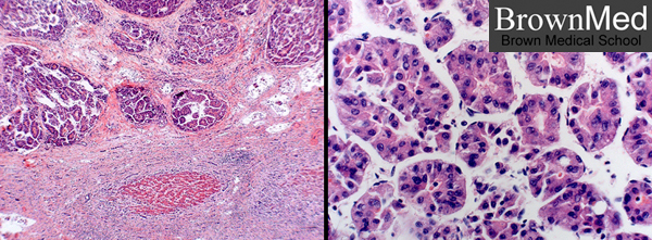
Hepatocellular carcinoma
The lower portion of the low magnification photo on the left shows a residual island of hepatocytes in an area of infiltrating
carcinoma, lymphocyte infiltration, and desmoplasia. The right higher magnification photo shows a moderately-
differentiated pseudoglandular pattern in this case. Well-differentiated HCC would present more clearly recognized
hepatocytes. In some cases of HCC, the cells are anaplastic. Cirrhosis of various causes is frequently a forerunner of HCC.