Bronchopulmonary disease
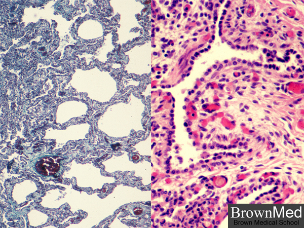
Microscopically, BPD shows fibrosis in interstitium of septal walls and elsewhere as seen on the left
with a trichrome stain. On the right, one sees hyperplasia of cuboidal epithelium lining the airspaces.
1 minute clinical correlation